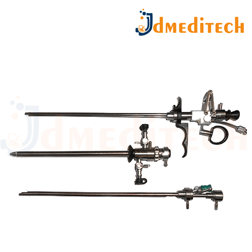
A Laser TURP Resectoscope Set is a specialized surgical tool used in Laser Transurethral Resection of the Prostate (Laser TURP) for treating benign prostatic hyperplasia (BPH). It combines a resectoscope with laser technology to remove prostate tissue.
Features:
Laser fiber for precise cutting and vaporization of prostate tissue
Resectoscope with a working channel for laser fiber and other surgical instruments
High-quality, sterilizable components
Adjustable laser settings for different tissue types and conditions
Clear visualization through camera coupling and light source
Uses:
Used in Laser TURP procedures to treat BPH by removing excess prostate tissue
Provides precise tissue removal with minimal bleeding
Offers a minimally invasive approach for improved recovery time and reduced risk of complications
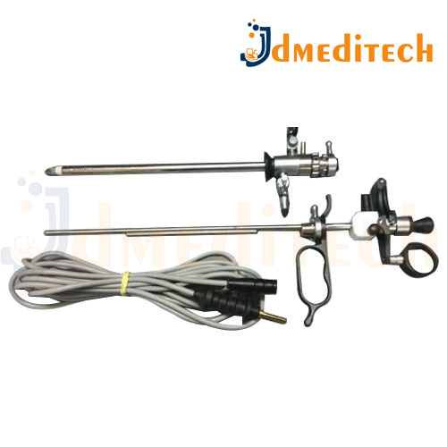
A Bipolar TURP Resectoscope Set is a specialized surgical instrument set used in Bipolar Transurethral Resection of the Prostate (TURP) to remove prostate tissue, designed with bipolar technology for safer and more efficient surgeries.
Features:
Bipolar technology for cutting and coagulating tissue simultaneously
Sterile, high-quality materials for durability
Resectoscope with a working channel for tools like electrodes and laser fibers
Adjustable settings for power and cutting depths
Smaller, more precise instruments for better control
Uses:
Used in TURP surgery to treat benign prostatic hyperplasia (BPH)
Minimizes risk of complications like TURP syndrome due to safer use of saline as the conductive medium
Provides efficient tissue removal while controlling bleeding and minimizing tissue damage
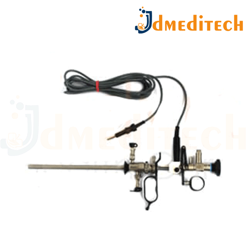
A Monopolar TURP Resectoscope Set is a complete surgical instrument set used for Transurethral Resection of the Prostate (TURP) using monopolar electrosurgery to treat BPH (Benign Prostatic Hyperplasia).
Features:
Includes:
Outer sheath and inner sheath
Working element (monopolar)
Monopolar loop electrode
Obturator and visual obturator
Compatible cable and telescope
Requires non-conductive irrigation fluid (e.g., glycine)
Enables precise tissue resection and coagulation
Designed for urethral access and safety
Uses:
Used in TURP surgery to resect prostatic tissue in patients with enlarged prostate
Ideal for cutting and coagulating using monopolar electrosurgical current
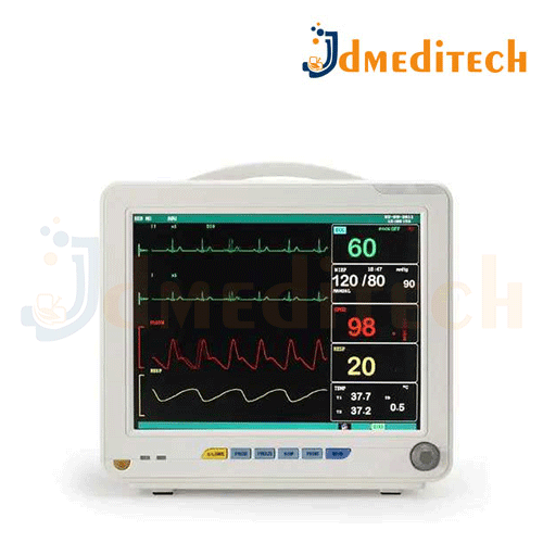
A Medical Monitor is a specialized display device used in healthcare settings to monitor and display real-time vital signs, medical images, or procedural data for patient care and surgical procedures.
Key Features:
High-resolution display for clear viewing of patient data and imaging
Real-time monitoring of vital signs (e.g., heart rate, oxygen levels, blood pressure)
Multi-input capability to connect with different medical devices (ECG, pulse oximeters, etc.)
Touchscreen or button controls for ease of use
Portable or stationary depending on the application
Compatibility with imaging devices (e.g., ultrasound, X-ray, endoscopy)
Alarm system to alert medical staff of abnormal readings
Uses:
Monitoring patient vitals during surgery or intensive care
Displaying diagnostic images during medical procedures (e.g., endoscopy, ultrasound)
Data storage and analysis for patient monitoring and review
Training and educational use in medical environments
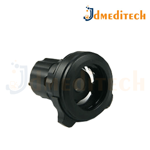
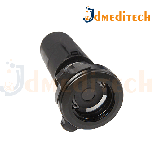
A Camera Coupler is a device used to connect a surgical camera to an endoscope or other medical instruments during minimally invasive procedures, ensuring proper alignment and transfer of images to a monitor.
Features:
Universal compatibility with various endoscopes and cameras
Durable and sterile materials for safe use
Precision alignment for clear image transmission
Quick and secure attachment to the camera and endoscope
Ergonomically designed for ease of use in surgery
Uses:
Used in endoscopic surgeries (e.g., urology, gastrointestinal, laparoscopy)
Ensures real-time visualization of the surgical site on a monitor
Facilitates minimally invasive procedures, allowing the surgeon to see and guide instruments with precision
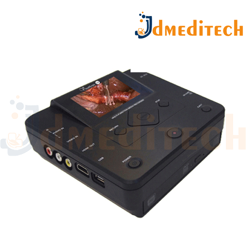
A Video Recorder in medical settings is used to capture, store, and display video images from endoscopic procedures, such as cystoscopies or laparoscopies, for documentation and analysis.
Key Features:
High-definition video capture for clear imaging
Storage capability for recording procedures
Connects to endoscopic cameras for live feed display
Playback function for reviewing recorded procedures
Compatible with various medical imaging systems
Digital or analog options
Uses:
Documenting surgical procedures for patient records
Reviewing and analyzing diagnostic images
Training and educational purposes in medical fields
Telemedicine for remote consultations or reviews
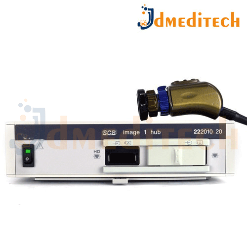
The Karl Storz Camera System is a state-of-the-art endoscopic imaging system used in minimally invasive surgeries such as laparoscopy, arthroscopy, urology, gynecology, ENT, and more. It delivers high-definition (HD) and 4K video for real-time visualization of internal organs, helping surgeons operate with precision, safety, and efficiency.
Key Features of Karl Storz Camera System:
HD and 4K Resolution Imaging – Provides ultra-clear and detailed visuals for accurate diagnosis and treatment.
IMAGE1 S™ and RUBINA™ Technology – Advanced platforms offering enhanced image processing, 3D, and NIR/ICG fluorescence imaging.
Integrated Light Source (LED/Xenon) – Offers strong, focused illumination for deep tissue visibility during procedures.
Autofocus & Digital Zoom – Enables better viewing control without manual adjustments.
Modular & Scalable Design – Compatible with a wide range of rigid and flexible endoscopes and easily upgradeable.
Recording & Documentation Features – Allows surgeons to capture videos and images for training, reporting, and analysis.
Specialty Support – Ideal for general surgery, urology, gynecology, ENT, orthopedics, and more.
Used In:
General & Laparoscopic Surgery
Urology (TURP, PCNL, Cystoscopy)
Gynecology (Hysteroscopy, Laparoscopy)
ENT (Sinus, Ear, Throat surgeries)
Orthopedics (Arthroscopy)
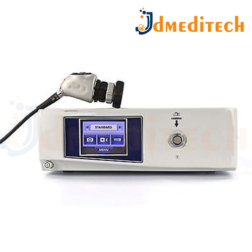
A Stryker Camera System is a high-performance medical video imaging system used during minimally invasive surgeries such as laparoscopy, arthroscopy, cystoscopy, and ENT procedures. Manufactured by Stryker Corporation, a leader in medical technology, this system provides ultra-clear, high-definition (HD, 4K, or even 8K) visuals to assist surgeons in real-time visualization inside the body.
Key Features of a Stryker Camera System:
HD/4K/8K Resolution – Delivers crystal-clear images with enhanced detail and depth.
Advanced Camera Control Unit (CCU) – Optimizes image clarity, brightness, and contrast during procedures.
Integrated LED or Xenon Light Source – Provides bright, focused illumination for deep tissue visibility.
Autofocus & Zoom Capabilities – Ensures precise visualization of anatomical structures.
Image & Video Recording – Supports documentation, review, and teaching.
Compatibility with Endoscopes – Works with a variety of rigid and flexible scopes across specialties.
Digital Output to Surgical Monitors – Real-time video display for entire surgical teams.
Used In:
Laparoscopic Surgery
Gynecology
Orthopedic Arthroscopy
Urology (e.g., TURP, cystoscopy)
General Surgery
ENT (Ear, Nose, Throat) procedures
A Wireless Camera is a digital video camera that transmits audio and video signals to a receiver or display device using Wi-Fi or radio frequency, rather than cables. In medical and surgical settings, wireless cameras are used to capture and stream live procedures without the clutter of wires, enhancing mobility, safety, and efficiency in the operating room.
Key Features of a Wireless Camera:
Wireless Connectivity – Eliminates cables, reducing clutter and improving mobility.
HD/Full HD/4K Resolution – Produces sharp and clear images for accurate observation or recording.
Battery Operated or Rechargeable – Allows portable and flexible use in different environments.
Real-Time Streaming – Live video transmission to monitors, tablets, or cloud storage.
Video Recording Option – Can store data locally (SD card) or on network/cloud systems.
Compact & Lightweight – Easy to mount on instruments, walls, or wearable headgear.
App/Software Support – Often controllable via smartphone or PC apps for remote operation.
Used In:
Medical field (surgery, endoscopy, dental, ENT)
Security & surveillance
Telemedicine and online consultation
Industrial equipment monitoring
Veterinary and mobile clinics
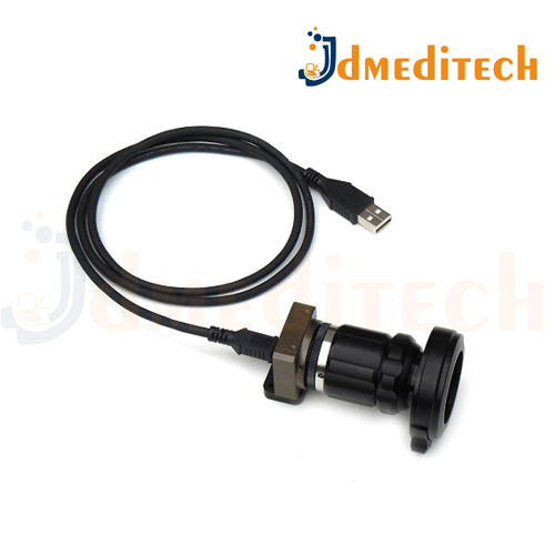
A USB Camera is a compact digital camera that connects to a computer or other devices via a USB port to capture, transmit, and display images or video.
Features:
High-resolution imaging for clear visuals
Plug-and-play functionality, no additional drivers required
Adjustable focus and zoom features in some models
Compatible with various software for video streaming or image capture
Often used with medical instruments like endoscopes, microscopes, or cameras for medical imaging
Use:
Commonly used in medical imaging for visualizing internal organs during surgeries or diagnostic procedures (e.g., endoscopy, ureteroscopy)
Streaming and recording of surgical or diagnostic procedures
Used for telemedicine or remote consultations in healthcare settings